Brain research on non-human primates – huge suffering for monkeys, no benefit for humans
Background
Non-human primates, mostly macaques, are still used for torturous brain experiments in Germany in basic research. Those experiments have no relevance for the research and treatment of human diseases, especially since diseases such as dementia, Alzheimer's, or Parkinson's do not occur naturally in those animals. Nevertheless, researchers justify their work primarily by stating that their findings about certain processes in the monkey brain could possibly be used in the future to understand human diseases affecting the brain. However, that apparent goal is not the specific aim. And that is not even the only reason that speaks against the cruel monkey brain research.
In the following, we exemplarily discuss some central aspects that show the scientific irrelevance of primate brain research for human health. We explain the different aspects of the animals' severe suffering and the lacking significance of the results from basic research. In addition, we demonstrate modern brain research methods that are relevant to humans and do not involve animal suffering. The focus here is on macaque monkeys. This genus includes rhesus monkeys (Macaca mulatta) and long-tailed macaques, also called Javanese monkeys (Macaca fascicularis). In brain research, rhesus monkeys are used almost exclusively, while long-tailed macaques suffer and die mainly in the field of toxicity testing.
Clear species differences in brain structures and functions between monkeys and humans
As humans are primates too, at first sight it seems obvious to use non-human primates as so called "model organisms” for basic research on how the human brain works. However, what should already be ethically completely out of question due to the close relationship and thus similar sentience, is also not a feasible way to draw conclusions about the human brain. This is due to species differences, that have already been widely established. Furthermore, there are now numerous human-relevant methods that can be used to specifically study human diseases.
The number of studies using imaging techniques and painless, non-invasive brain imaging in humans is growing. The discrepancies between data obtained in humans and non-human primates are becoming increasingly apparent. From numerous publications published in peer-reviewed journals, we present just a few differences between humans and non-human primates, with a focus on macaques.
- Primate brains are known to differ "both in aspects of structural detail and in general size." Differences in brain size are evident not only in differences in cell number, but also in the arrangement of cells and their connectivity (1).
- The human cerebral cortex has a surface area ten times larger than the macaque cortex and plays a key role in human cognition (2).
- There are major differences in gyrification (formation of furrows and convolutions) between the macaque and the human brain (3).
- The developmental time of brain cells (neurons) differs between species as it is subject to different processes: for example, these processes take 100 days in humans and only 60 days in macaques (4).
- A comparison of cell connections and linkages in human brains with those in macaque brains shows that almost half (45%) differ substantially (5).
Nevertheless, macaques and other non-human primates are continuously used as a so-called model for humans, especially for research on the processing of visual, auditory, and acoustic stimuli in the brain. This is increasingly being questioned from the scientific community as well:
For example, a review of numerous studies on human face recognition (6) urges caution in drawing direct conclusions between species without sufficient similarities in behavior and anatomical-functional landmarks, because there are numerous differences:
- Unlike humans, macaques and other monkeys are unlikely to be able to recognize individual conspecifics by their faces. There is no evidence that they rely on qualitatively similar representations like humans (e.g., fine-grained holistic representations of faces that allow discrimination at a glance) (6).
- In the visual cortex, the primary visual cortex is the largest area in both humans and macaques, but as a proportion of the total cerebral cortex it occupies a significantly larger area in macaques (10 percent vs. 3 percent) (6).
- Area V2 (shape and contour recognition) is proportionally much smaller in humans than in the macaque cortex (2).
- In contrast, the temporal lobe (important in humans for hearing, vision, and memory) is significantly smaller in macaques than in humans, even relative to body size (7).
- The rhesus monkey's face processing system, located in the cerebral cortex, lacks crucial aspects for human competence in recognizing individual faces (6).
- The structure of the visual cortex differs significantly between humans and macaques (8-11).
Numerous studies in the field of monkey brain research work with the processing of movement stimuli. Although motion stimuli address broadly similar regions in the cerebral cortex in humans and macaques, there are striking functional differences (review article: 12):
- Motion processing is much more prominent in the human brain with respect to the coordination of visual perception and motor activity (e.g., coordination of eye movement with goal-directed grasping) than in monkeys (12).
- The number of motion-sensitive regions is reduced in monkeys compared to humans (13).
- Several regions in the temporal lobe of humans are significantly more sensitive to motion stimuli than in monkeys (12).
- The involvement of the parietal lobe in movement processing is more limited in monkeys (14)
- It is suggested that movement processing became particularly essential for humans when they began to move across wide open plains in search of food, whereas apes primarily forage in trees (12).
The motor speech center in the brain of nonhuman primates (areas 44 and 45) has been described as comparable to Broca's region in humans (speech production and speech perception). However, there are significant differences, which are seen as causative for the much more complex and far-reaching meaning of language in humans:
- The human central nervous system has the densest intercellular neural network compared to apes and by a considerable margin in comparison to macaques, too. Thus, the neurons of the different brain regions are much better connected in humans than in other species (15).
- In this area, the tissue architecture of humans differs considerably from that in non-human primates, particularly gibbons and macaques (15).
The frontal lobe in humans plays an important role in language (16) and cognitive control of behavior (17-21). Key features of its structure appear to be comparable in humans and macaques (22). However, there are authoritative species differences that illustrate that apparent structural similarity has little explanatory power on behavior and cognitive performance between species (23):
- Heard information is of much higher importance to humans than to macaques. The latter are less able to remember auditory information than humans (24), whereas they can easily learn to use visual information (25).
- The anterior part of the cerebral cortex in humans contains a region not found in macaques (23).
- The linkage of the various brain regions responsible for processing auditory information with other brain regions differs greatly between humans and macaques (23,26).
Already due to the - not conclusive - differences between the brains of humans and non-human primates presented here, the usefulness of monkey brain research is more than questionable from a purely scientific point of view. Many of these comparisons between the brains of monkeys and humans originate from monkey brain research itself – which even makes it more puzzling that non-human primates continue to be used as "model organisms" for humans. Due to these differences, the "findings" obtained in this way simply cannot be transferred to humans, and thus cannot provide any help for sick people.
Lack of validity of the results of monkey brain research
At best, research on monkey brains allows statements about specific aspects of the processes in the brain of the studied monkey species. Transferring these findings to humans is pure speculation anyway, and especially since not even healthy animals are studied, but severely damaged ones. Numerous voices - including those of researchers themselves - emphasize that results obtained from such stressed and sick animals are not representative.
This is because using stressed and suffering animals in research compromises the validity, reliability, and reproducibility (repeatability) of the "findings" obtained. Studies show that suboptimal housing, removal from the social group, and switching to different housing (even if it is social) is also associated with several physiological changes indicative of stress in the monkeys (27 - 30). Stress affects the brain in multiple ways, including highly cognitive processes (31,32). The effects of stress are not limited to the brain but extend to the whole organism as well as to an animal’s behavior (33). These changes limit the validity of research findings - after all, abnormal physiology often underlies observable abnormal behavior (34 - 39).
However, even the absence of behavioral indicators of stress do not necessarily show that there are no physiological changes. Studies of restrained animals in primate chairs (40, 41) concluded that monkeys first exhibited anxiety-related behaviors but promptly appeared calm. Their anxiety behaviors ceased with repeated exposure to the laboratory situation and levels of the stress hormone cortisol returned to baseline levels. However, the researchers don´t attribute this to a lowering of stress levels, but rather to an attenuation of the cortisol response. This also occurs in humans under continuous stress with numerous negative health effects.
In this context, the "normal" functionality of the monkeys' brains may be strongly affected and permanently altered by the prolonged stress and pain (unavoidable in brain research) (42). Changes in the body’s water balance, which occurs when animals are suffering from thirst during experimental periods, can also have an impact on cellular function (43, 44). The anesthesia often used for studies with noninvasive imaging techniques such as fMRI limits the validity not only because the monkeys are not awake during these procedures, but also because anesthetics can alter responses and confound results (42, 45).

The validity of research on severely stressed animals is more than questionable.
The suffering of monkeys for basic research
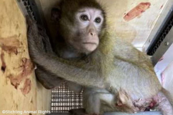
Macaques are most commonly "used" at institutes conducting brain research. The suffering of the animals is not limited to the experiment. The monkeys are torn away from their mothers and families at a way too young age. They spent prolonged time all alone during quarantine, and are shipped in dark boxes under adverse conditions for days - often originating from Asia or Mauritius - to end up in European or US laboratories. Investigations published in 2024 revealed shocking deficiencies in the transport of long-tailed macaques, which were shipped to laboratories worldwide via Brussels Airport (46).

Non-human primates are being transported in small wooden crates from Asia or Mauritius via the hub in Brussels into European laboartories. More images by Stichting Animal Rights >>
In some cases, the animals come from breeding farms or even from the wild (47- 49). Particularly alarming: Since 2022, long-tailed macaques have been classified as endangered species by the International Union for Conservation of Nature (IUCN) (50). On the way to the laboratories, there are usually intermediate stops via further institutions. Those institutions usually also breed monkeys in the countries themselves but cannot meet the demand - therefore they buy monkeys from Asia. The traumatized young animals spend some time here, before they are sold on (51, 52). And again, they are torn away from their (new) group to end up in a laboratory.
After some time of acclimation in the laboratories, the young monkeys are "trained" to climb into the primate chair, allow themselves to be restrained, and perform certain tasks on the screen under water deprivation. Due to the life-threatening thirst, they have no other option than to surrender to their fate. This "training" alone can last for many months. If the animals behave according to the researcher's wishes, the first operations take place.

Baby monkeys are often separated from their mother and family far too early.
The brain surgeries performed are highly invasive. The animals are implanted with measuring devices and head posts on and in the skull. The skull bone and bone skin are perforated in the process. During the measurements, electrodes are inserted into the brain regions relevant to the research question. Each monkey is usually "used" several times, often over a period of years, for different questions. This means that the measuring instruments also have to be moved and that there is a further perforation of the skull at a new location. In addition, there are holes for the screws with which the instruments are attached to the skull. The respective "training" in order for the animals to react to the right stimuli is also repeated several times as a result.
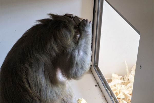
For data recording, the monkeys are locked in a primate chair and their heads are screwed to a mount so that they are completely unable to move. Otherwise, measurement would not be precise. The animals sit in front of the screen for hours each day in this way and have to solve tasks while their brain waves are measured. In order for the animals to solve the tasks and cooperate, they are only given fluid during these trials, and when they solve the task correctly. This is their only way to quench their thirst, because away from the tests or training, the animals do not receive any fluids during their "work week."
The animals are thus forced into considerable suffering (the fixation in the primate chair) to escape a potentially life-threatening situation – dehydration. Animal experimenters have been presenting this circumstance as harmless for decades, even talking about positive reinforcement. But in that case, it would not be necessary to create a deficiency (dehydration), because the monkeys would participate in the experiment voluntarily, i.e. even without thirst, for the reward (e.g., desirable but not vital food) received. However, the fact that a life-threatening deprivation is needed for the animals to cooperate is the best illustration that they perceive the experiments as suffering.

Non-human primates with devices implanted on their skulls.
A 2009 veterinary pathology report conducted on dead monkeys from brain research at the Max Planck Institute for Biological Cybernetics in Tübingen, Germany, found severe suffering. In the monkey Jara, these included multiple drillholes in the skull bone, circular dead skin and inflammation of the meninges, a severed masseter muscle, puncture wounds to the brain, and severe traumatic brain injury. Necropsy revealed the cause of death as "chronic severe traumatic brain injury, neurogenic shock with presumed severe pain." Internal correspondence indicates that "the operation was in accordance with the normal procedure at the Institute" and that "the surgeon was one of the most competent for neuroimplants at the MPI" (53).
If the monkeys live in groups with animals that get along well, this can alleviate their suffering at least during the test-free period. But the animals are regularly taken out of the group to participate in the tests - the group structures are thus shaken anew each time. A returning animal must always fear for its status. This results in a considerable stress factor. If the animals, which are often socially damaged at an early age, do not (or no longer) get along with other group members, they often spend the rest of their lives in solitude . This alone is a considerable torment and severe suffering for the social animals. Sometimes "trained" monkeys are also given to other laboratories, where the suffering continues. Under usual protocol, however, the macaques are killed at the end of the test series - this can be the case after many years - as it is stated that brain slices are needed for an evaluation of the data collected in advance.
In 2024, information and undercover recorded footage from the Ernst Strüngmann Institute (ESI) in Frankfurt were made public, highlighting the suffering of the animals and, in particular, the duration of their suffering in brain research. Some of the monkeys were used in experiments for between 10 and 20 years (54).

Over 20 years with devices implanted on his skull: rhesus monkey Homer at the Ernst Strüngman Institute Frankfurt. Credit: SOKO Tierschutz
Possibilities of animal-free, human-based brain research
If fundamental knowledge about the functions of the human brain is to be gained in order to understand and cure human diseases such as Alzheimer's or Parkinson's disease, numerous non-invasive, animal-free and human-based research methods are already available today. Studies of so-called brain lesions (functional failures of individual brain regions) in humans made a significant contribution to gaining knowledge of brain functions early on (55). Moreover, since as early as the 1950s, brain research has worked with voluntary and comprehensively informed human patients (56). Since then, both invasive and noninvasive ethically justifiable human studies have contributed significantly to neuroscience knowledge. Studies examining human brain tissue (obtained from brain surgery or from deceased humans) remain important today in providing insights into brain anatomy.
Electrical stimulation and more invasive recording of brain activity can also be performed during brain surgery. These are cases where patients have to undergo such procedures for medical reasons and are informed in detail in advance about the additional experiment, its implications and possible risks. Thus, unlike animals used, they may give informed consent to their participation. These tests on humans also offer the great advantage that through direct communication with the patient, the results can be interpreted more easily. Head electrodes, as well as various imaging techniques such as fMRI (functional magnetic resonance imaging) or fNIRS (functional near-infrared spectography) are used to study brain activity in awake volunteers. Mathematical models work with data from population studies and have, for example, already provided important insights for a therapy development for multiple sclerosis (57). Newer methods such as 3D printing can be used to accurately map damage in patients' brains, leading to a better understanding of disease progression and thus improvements in therapies for a wide range of neurodegenerative diseases.

Various imaging techniques can be used to analyse the brain activity of volunteers - which, in contrast to animal testing, provides meaningful results.
Also particularly promising are the so-called mini brains - millimeter-sized, three-dimensional structures made of human cells that mimic many anatomical and functional properties of the human brain. Compared to traditional 2D cell cultures, Mini Brains can have multiple layers of different, interconnected cell types. Thus, they offer scientists the opportunity to study brain processes, diseases, and therapies in a complex, human-relevant, and person-specific system without performing invasive procedures on patients (57).
Conclusion
In summary, monkey brain research shows only one thing with certainty: how the brain of the studied traumatized, sick, and severely stressed and suffering monkey of a specific sex and species reacts to certain stimuli at a specific moment - without implications for other monkeys, let alone humans. This is because both brain structure and the functioning of different brain areas differ too much between monkeys and humans. The results from monkey brain research are simply not transferable to humans.
Even if the cruel monkey brain experiments had a benefit for humans - which is explicitly not the case - they should not be allowed to be carried out because it is ethically impermissible to torture other animals and especially our closest relatives in this way. Monkeys have been suffering since childhood when they are snatched from their families to suffer the horrific ordeals in the laboratory. Monkeys, despite the many differences in brain functionality presented, are very similar to humans not only in terms of their intelligence, but also in their emotional world. They have a rich and complex social and emotional life, feeling joy, sadness, envy, and jealousy, and displaying a high degree of empathy and also selflessness - traits that until a few years ago were only attributed to humans. But the findings from the cruel experiments are not even of any help to sick human patients.
Nevertheless, millions of research dollars are being spent on research that is of no benefit to humans - tax dollars that could be used to gain actual applicable knowledge for human patients through human-oriented brain research using the numerous human-based, non-animal methods available.
17 December 2024
Dr Corina Gericke, DVM
References
- Rilling J.K. Human and nonhuman primate brains: are they allometrically scaled versions of the same design? Evolutionary Anthropology: Issues, News, and Reviews 2006; 15(2): 65-77
- Van Essen D.C. A population-average, landmark-and surface-based (PALS) atlas of human cerebral cortex. Neuroimage 2005; 28(3): 635-662
- Zilles K. et al. Development of cortical folding during evolution and ontogeny. Trends in neurosciences 2013; 36(5): 275-284
- Geschwind D.H. et al. Cortical evolution: judge the brain by its cover. Neuron 2013; 80(3): 633-647
- Goulas A. et al. Comparative analysis of the macroscale structural connectivity in the macaque and human brain. PLoS computational biology 2014; 10(3): e1003529
- Rossion B. et al. What can we learn about human individual face recognition from experimental studies in monkeys? Vision research 2019; 157: 142-158
- Rilling J.K. et al. A quantitative morphometric comparative analysis of the primate temporal lobe. Journal of Human Evolution 2002; 42(5): 505-533
- Barton J.J. Structure and function in acquired prosopagnosia: lessons from a series of 10 patients with brain damage. Journal of neuropsychology 2008; 2(1): 197-225
- Meadows J.C. The anatomical basis of prosopagnosia. Journal of Neurology, Neurosurgery & Psychiatry 1974; 37(5): 489-501
- Rossion B. Understanding face perception by means of prosopagnosia and neuroimaging. Frontiers in bioscience 2014; 6 (258): e307
- Schira M.M. et al. Brain mapping: the (un)folding of striate cortex. Current Biology 2012; 22 (24): 1051-1053
- Orban G.A. et al. Similarities and differences in motion processing between the human and macaque brain: evidence from fMRI. Neuropsychologia 2003; 41(13): 1757-1768
- Simon O. et al. Topographical layout of hand, eye, calculation, and language-related areas in the human parietal lobe. Neuron 2002; 33(3): 475-487
- Tootell R.B. et al. Functional analysis of V3A and related areas in human visual cortex. Journal of Neuroscience 1997; 17(18): 7060-7078
- Palomero-Gallagher N. et al. Differences in cytoarchitecture of Broca's region between human, ape and macaque brains. Cortex 2019; 118: 132-153
- Friederici A.D. et al. The language network. Current opinion in neurobiology 2013; 23(2): 250-254
- Aron A.R. The neural basis of inhibition in cognitive control. The neuroscientist 2007; 13(3): 214-228
- Brass M. et al. The role of the inferior frontal junction area in cognitive control. Trends in cognitive sciences 2005; 9(7): 314-316
- Dosenbach N.U. et al. A core system for the implementation of task sets. Neuron 2006; 50(5): 799-812
- Higo T. et al. Distributed and causal influence of frontal operculum in task control. Proceedings of the National Academy of Sciences 2011; 108(10): 4230-4235
- Neubert F.X. et al. Cortical and subcortical interactions during action reprogramming and their related white matter pathways. Proceedings of the National Academy of Sciences 2010; 107(30): 13240-13245
- Petrides M. et al. Comparative cytoarchitectonic analysis of the human and the macaque ventrolateral prefrontal cortex and corticocortical connection patterns in the monkey. European Journal of Neuroscience 2002; 16(2): 291-310
- Neubert F.X. et al. Comparison of human ventral frontal cortex areas for cognitive control and language with areas in monkey frontal cortex. Neuron 2014; 81(3): 700-713
- Scott B.H. et al. Monkeys have a limited form of short-term memory in audition. Proceedings of the National Academy of Sciences 2012; 109(30): 12237-12241
- Gaffan D. et al. Auditory-visual associations, hemispheric specialization and temporal-frontal interaction in the rhesus monkey. Brain 1991; 114(5): 2133-2144
- Rudebeck P. H. et al.). A role for the macaque anterior cingulate gyrus in social valuation. Science 2006; 313 (5791): 1310-1312
- Gust D.A. et al. Effect of a preferred companion in modulating stress in adult female rhesus monkeys. Physiology & behavior 1994; 55(4): 681-684
- Baker K.C. et al. Pair housing for female longtailed and rhesus macaques in the laboratory: behavior in protected contact versus full contact. Journal of Applied Animal Welfare Science 2012; 15(2): 126-143
- Capitanio J.P. et al. Social instability and immunity in rhesus monkeys: the role of the sympathetic nervous system. Philosophical Transactions of the Royal Society B: Biological Sciences 2015; 370(1669): 20140104
- Garner J.P. Stereotypies and other abnormal repetitive behaviors: potential impact on validity, reliability, and replicability of scientific outcomes. ILAR journal 2005; 46(2): 106-117
- McEwen B.S. et al. Mechanisms of stress in the brain. Nature neuroscience 2015; 18(10): 1353-1363
- Teicher M.H. et al. The effects of childhood maltreatment on brain structure, function and connectivity. Nature reviews neuroscience 2016; 17(10): 652-666
- McEwen B.S. Stress, adaptation, and disease: Allostasis and allostatic load. Annals of the New York academy of sciences 1998; 840(1): 33-44
- Suomi S.J. et al. Depressive behavior in adult monkeys following separation from family environment. Journal of abnormal psychology 1975; 84(5): 576
- Cohen et al. Chronic social stress, affiliation, and cellular immune response in nonhuman primates. Psychological Science 1992; 3(5): 301-305
- Gordon T.P. et al. Social separation and reunion affects immune system in juvenile rhesus monkeys. Physiology & behavior 1992; 51(3): 467-472
- Gust D.A. et al. Removal from natal social group to peer housing affects cortisol levels and absolute numbers of T cell subsets in juvenile rhesus monkeys. Brain, Behavior, and Immunity 1992; 6(2): 189-199
- Gust D.A. Effect of a preferred companion in modulating stress in adult female rhesus monkeys. Physiology & behavior 1994; 55(4): 681-684
- Capitanio J.P. et al. Individual differences in peripheral blood immunological and hormonal measures in adult male rhesus macaques (Macaca mulatta): evidence for temporal and situational consistency. American Journal of Primatology 1998; 44(1): 29-41
- Golub M.S. et al. Adaptation of pregnant rhesus monkeys to short-term chair restraint. Laboratory Animal Science 1986; 36(5): 507-511
- Ruys J.D. et al. Behavioral and physiological adaptation to repeated chair restraint in rhesus macaques 2004. Physiology & behavior;82(2-3): 205-213
- Martin C. Contributions and complexities from the use of in vivo animal models to improve understanding of human neuroimaging signals. Frontiers in neuroscience 2014; 8: 211
- Machaalani R. et al. Effects of changes in energy homeostasis and exposure of noxious insults on the expression of orexin (hypocretin) and its receptors in the brain. Brain research 2013; 1526: 102-122
- Kallaras C. et al. Intracerebroventricular administration of atrial natriuretic peptide prevents increase of plasma ADH, aldosterone and corticosterone levels in restrained conscious dehydrated rabbits. Journal of endocrinological investigation 2004; 27: 844-853
- Masamoto K. et al. Anesthesia and the quantitative evaluation of neurovascular coupling. Journal of Cerebral Blood Flow & Metabolism 2012; 32(7): 1233-1247
- Doctors Against Animal Experiments: Appalling conditions uncovered during monkey transports to laboratories. Press release, 15.11.2024
- Southeastasiaglobe: Cambodias monkey farms corruption taints wildlife trait. 04/2023 [accessed on 05.08.2023]
- The Guardian: Thailands missing monkeys – why did hundreds of rhesus macaques vanish from a temple. 02/2023 [accessed on 05.08.2023]
- Capital: Laboraffen aus China werden knapp. 16.10.2022 [accessed on 05.08.2023]
- Ärzte gegen Tierversuche: Häufigste im Tierversuch verwendete Affenart gilt als vom Aussterben bedroht. Pressemitteilung vom 20.09.2023
- LSCV: Primatenzucht in Mauritius. 25.02.2019 [accessed on 05.08.2023]
- One Voice: Silabe, an establishment within the University of Strasbourg, at the heart of the international trade in monkeys for animal experimentation. 03/2021 [accessed on 05.08.2023]
- Ärzte gegen Tierversuche: Neue Beweise: So leiden Affen in Deutschland in der Hirnforschung. [accessed on 05.08.2023]
- Doctors Against Animal Experiments: Close ESI Frankfurt - Freedom for the monkeys! Save Gandalf! [accessed on 01.12.2024]
- Damasio et al. The return of Phineas Gage: clues about the brain from the skull of a famous patient. Science 1994; 264(5162): 1102-1105
- Ward A.A. et al. The electrical activity of single units in the cerebralcortex of man. Electroencephalography and Clinical Neurophysiology 1955; 7: 135-136
- Kotelnikova E. et al. Dynamics and heterogeneity of brain damage in multiple sclerosis. PLoS Computational Biology 2017; 13(10):e1005757
- Doctors Against Animal Experiments: Mini brains: innovative, human-relevant brain research [accessed on 05.08.2023]
